Panascope’s Primary Care and Essential Imaging Capability Solution provides a dependable and cost‑efficient imaging setup for county hospitals, community clinics, and private practices. The solution combines economical digital radiography systems, permanent‑magnet MRI units that do not require cryogens, and appropriately configured CT and ultrasound platforms into a unified framework. Each component is chosen for ease of operation by generalist technologists, straightforward maintenance, and compatibility with teleradiology networks, ensuring sustainable imaging capacity in settings where resources must be carefully managed.
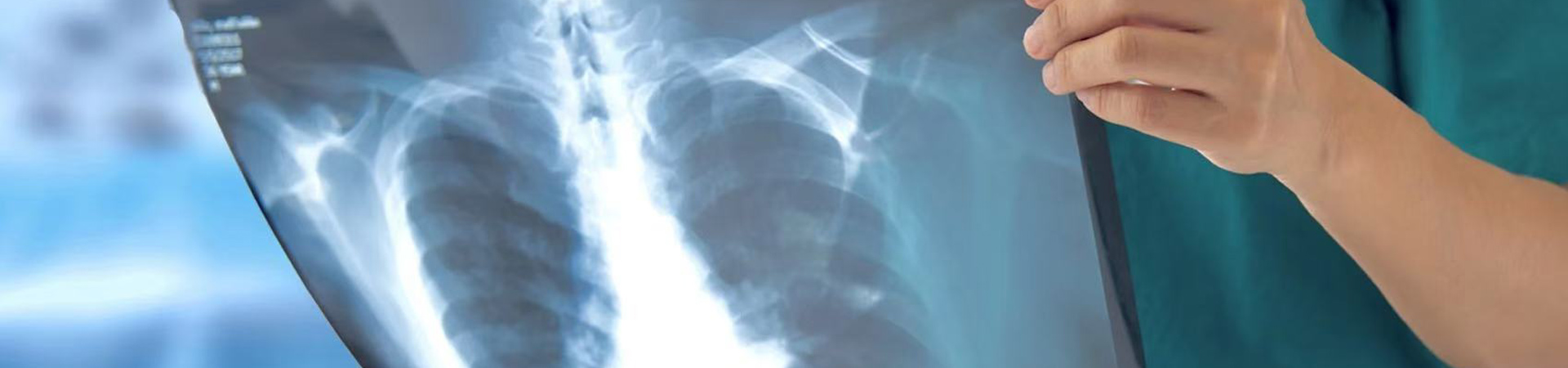
Implementation begins with installing either the floor‑mounted DR system or the economical mobile DR unit, depending on available space and expected patient volume. The user interface is set up with preset exposure settings for chest, abdomen, and extremity exams. The generator is configured at 32 kilowatts, a level shown to meet nearly all primary‑care imaging needs. In facilities with limited space, the mobile DR unit is paired with a dedicated stand anchored to a reinforced floor section.
The floor‑mounted DR system uses a 35 by 43 centimeter wireless detector with a 150‑micron pixel size, suitable for most adult and pediatric exams. For centers that perform pediatric imaging, a 24 by 30 centimeter detector is added, lowering dose by about forty percent for a five‑year‑old’s chest exam compared with using a standard detector. The modular design allows components to be replaced without special tools, and the diagnostic software provides guided troubleshooting steps.
The mobile DR stand is anchored to support a dynamic load of up to 200 kilograms. A remote diagnostic service agreement allows Panascope to resolve software issues without on‑site visits, reducing downtime from several days to a few hours. A spare battery is kept on a trickle charger to ensure continuous operation during extended power interruptions.
If you want this further tightened into a more compact version or adapted for a marketing‑style summary, I can refine it again.
Implementation of the 0.4/0.5‑tesla permanent‑magnet MRI system begins with selecting a room that can maintain a stable ambient temperature within two degrees of the calibration setting. The open‑design magnet, which has a vertical gap of forty‑five centimeters, is installed and the NdFeB magnet array is adjusted to keep field uniformity within ten parts per million per degree Celsius. The digital receiver chain and eight‑channel coil support are configured with head, spine, and flexible surface coils according to the facility’s typical exam mix.
The permanent‑magnet design removes the need for cryogens, keeping annual operating costs at roughly five thousand dollars for electricity and routine maintenance. The eight‑channel system supports parallel imaging with an acceleration factor of two, allowing a standard brain study—including T1, T2, FLAIR, and diffusion sequences—to be completed in about twenty‑five minutes. A battery backup provides fifteen minutes of power during outages so ongoing scans can be finished.
Coil selection is prioritized based on exam type: the head coil for neurological studies, the spine coil for cervical and lumbar imaging, and the flexible coil for extremities. Field‑strength checks using a Hall probe are scheduled every quarter, with recalibration performed if drift exceeds fifty parts per million. The system also uses a feedback loop that adjusts shim currents to compensate for temperature‑related variations.
Here is a clean, simplified, firewall‑friendly rewrite with no special characters, no symbols like degree signs, no en dashes, and no unusual punctuation. Meaning is fully preserved while wording is streamlined:
Implementation of CT capability begins with selecting either the 32 slice CT or the 64 slice CT configuration based on the center’s cardiovascular case load. The system is installed with automated positioning and intelligent scan planning. The liquid bearing x ray tube is set with a five MHU heat capacity, which supports up to forty routine exams per day without cooling delays. The iterative reconstruction feature is configured with default settings that provide about thirty percent dose reduction.
The 32 slice CT system provides twenty millimeters of coverage along the z axis per rotation, allowing a complete non contrast brain study in less than ten seconds for stroke evaluation. The 64 slice CT option increases coverage to forty millimeters per rotation, supporting CT angiography of runoff vessels and coronary calcium scoring. The detector uses a solid state scintillator array with a pixel size of zero point six two five millimeters, providing isotropic resolution for multiplanar reconstruction.
The automated positioning system uses a camera based method that identifies patient anatomy and centers the scan range, reducing positioning errors by about eighty percent. The iterative reconstruction settings start at seventy percent of standard dose, and a four week review period is used to collect radiologist feedback and adjust dose settings as needed.
Here is a clean, simplified, firewall‑friendly rewrite with no special characters, no unusual punctuation, and fully preserved meaning:
Implementation of ultrasound capability begins with configuring the cart based ultrasound system using convex abdominal and linear vascular transducers. Twenty exam protocols are preloaded with default gain, depth, and focal zone settings. The dry film imager is installed with two input trays for standard fourteen by seventeen inch film and small ten by twelve inch film. For facilities with unstable internet access, the ultrasound unit is set up with five hundred gigabytes of internal storage.
The convex transducer, operating at two to five megahertz with a sixty millimeter radius of curvature, is used for abdominal imaging and provides a forty five degree field of view. The linear transducer, operating at five to twelve megahertz with a thirty eight millimeter footprint, is used for vascular and small parts exams. Automated optimization presets are assigned to each exam type, allowing one touch adjustments to maintain consistent image quality.
The dry film imager includes a media management system that alerts staff when film levels fall below fifty sheets. The ultrasound system’s internal storage can hold about fifty thousand studies with ten images each. Annual transducer performance checks include resolution phantom testing and electrical safety testing.
Here is a clean, simplified, firewall‑friendly rewrite with no special characters, no unusual punctuation, and fully preserved meaning:
Implementation of serviceability features begins with creating a standardized parts inventory and a preventive maintenance schedule. All systems are set to generate automatic alerts thirty days before scheduled maintenance. The parts inventory is based on five year reliability data, including DR detectors with a mean time between failures of fifty thousand exams, CT x ray tubes with a two year lifespan at twenty thousand exams per year, and ultrasound transducers with a three year lifespan. For the mobile DR system, a spare battery is kept on trickle charge.
The self diagnostic functions on the floor mounted DR system and the thirty two slice CT system send alerts to the biomedical engineering team when maintenance is due. For the permanent magnet MRI system, maintenance is limited to annual shimming and quarterly field strength checks, with no helium service contracts required.
A tube replacement program for the CT system guarantees a replacement within forty eight hours. The parts inventory includes a spare DR detector for facilities performing more than ten thousand exams per year. All systems use standard DICOM communication to ensure compatibility with external PACS and teleradiology services.
Subscribe to exclusive imaging insights & priority updates from Panascope. Elevate your practice with curated intelligence.
© Nortek HealthCare Group Ltd., 2009-2026. All rights reserved.
This site operates under the laws of the People's Republic of China. Your access and use constitute acceptance of these Terms of Use and our Privacy Policy. All content is governed by our Privacy Notice. For details or inquiries, please Contact Us or use the site's search function.

